DEXA Examinations estimate the amount of bone mineral content in specific areas of your body by utilizing two types of x-rays, both of which allow Radiologists to make an estimation of bone density by showing the difference between bone and soft tissue. The examination is safe, quick, and painless—you won’t require any injections, sedation, special diet, or other advance preparation.
Nearly 10 million Americans have osteoporosis—a bone disease that greatly increases the risk of bone fracture. Common risk factors for osteoporosis include:
DEXA Examinations are critical in both diagnosing and subsequently treating osteoporosis, as well as other conditions that cause bone loss. Talk to your doctor about your risk factors for osteoporosis, and the importance of a DEXA screening.
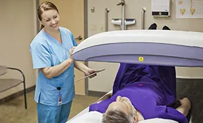
DEXA Examinations typically consist of a scan of the spine and a single hip. The exam itself takes about 20 minutes to perform. Our technician will ask you to lay still on a scan table and breathe normally while the scanner passes over the areas being evaluated. After the exam is complete, you are immediately able to return to your normal activities!
Your CareMedica provider will discuss the results with you and explain their significance. If you have lower-than-usual bone density, your provider may recommend treatments to help maintain strong bones, such as:
To schedule your DEXA Bone Density scan, please call (203) 672-2800 for our Connecticut offices and (561) 776-8890 for our Florida office. Patients can also schedule appointments through the Healow patient portals:


