An ultrasound, or sonography, is a diagnostic imaging technique used to visualize muscles, tendons, and many internal organs. It is commonly used to evaluate the size, structure, and any pathological lesions of the abdominal and pelvic organs, breast, thyroid glands, and heart, as well as the blood flow in arteries and veins.
A carotid ultrasounds is a safe and painless procedure that employs sound waves to assess the blood flow through the carotid arteries. Additionally, carotid ultrasounds evaluate the thickness of the carotid artery wall and detects any presence of clots.
Situated on each side of the neck, the carotid arteries play a crucial role in delivering blood from the heart to the brain. The primary purpose of a carotid ultrasound is to identify any blockages or narrowing in the carotid arteries, which can heighten the risk of stroke. By analyzing the test results, your CareMEDICA healthcare provider can devise an appropriate treatment plan to mitigate your stroke risk.
There is no injection or radiation exposure associated with ultrasound. Please note that although we offer this service, the tests are not done in-house.
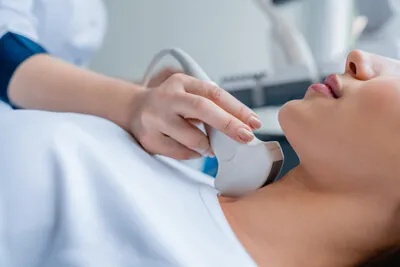
Depending on the type of study, an ultrasound typically takes between 15 and 30 minutes to complete. Our sonographer will help position you on a comfortable exam table and a clear gel, used as a conductor, will be applied to your skin. A hand-held device that sends and receives ultrasound waves called a transducer is moved back and forth over the area of your body being imaged.
These images are captured on a television-like monitor and transferred to film for our radiologist to review and interpret for your physician. After the exam is complete you will be able to return to your normal activities.
Exam Preparation:
An echocardiogram employs sound waves to generate images of the heart, offering insights into blood flow through its chambers and valves. This widely performed diagnostic tool enables healthcare providers to detect various heart conditions and diseases.
Alternate terms for an echocardiogram include:
Different echocardiogram variations are available, tailored to specific
testing purposes and individual health statuses. Some types of
echocardiograms can be conducted during exercise or pregnancy, depending
on the specific requirements of the situation.
There are different types of echocardiograms. The type of echocardiogram
you receive depends on the reason for the test and your overall
health. Some types of echocardiograms be done during exercise or
pregnancy. preparing for an echocardiogram requires no dietary
restrictions. Patients should anticipate the procedure to
take approximately 30-60 minutes.
Interested in being seen by a CareMEDICA provider? Call (203) 672-2800 for our Connecticut offices, and (561) 776-8890 for our Florida office. You can also schedule your appointment using our Healow portal:


